Discover the best moments of the olympics on a nanoscale
Replicating an elite athlete's performance at the Paris 2024 Olympics on a nanoscale? It can be done at the University of Twente's NanoLab!
A group of enthusiastic PhD students from MESA+ will replicate the most beautiful finish images of Dutch top athletes on the nanoscale during the Olympic Games - under the name Nanolympics.
REMCO EVENEPOEL - Time trial
Remco Evenepoel - Time trial | 27 July 2024, Olympics, Paris 2024
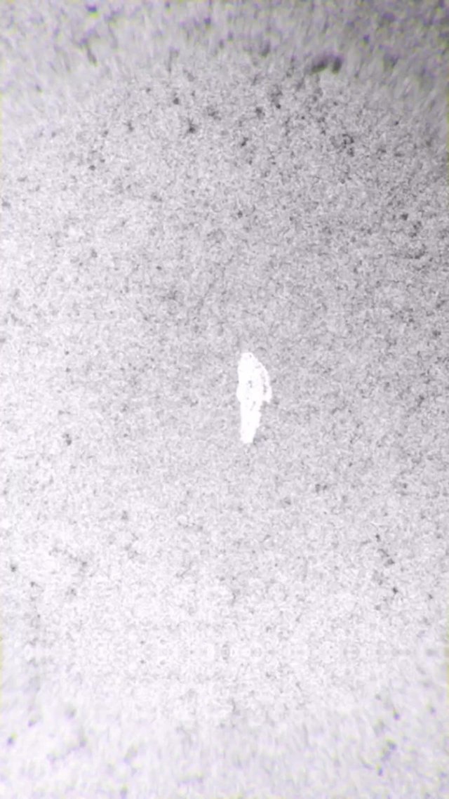
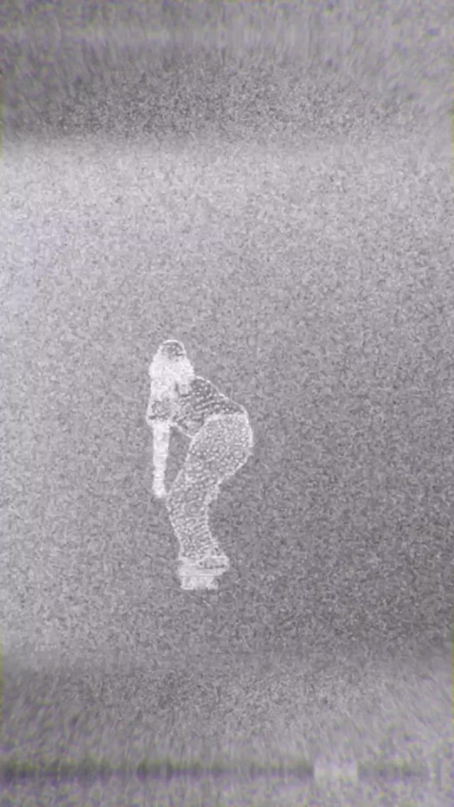
Keet Oldenbeuving - skateboarding
Keet Oldenbeuving - skateboarding | 27 July 2024, Olympics, Paris 2024
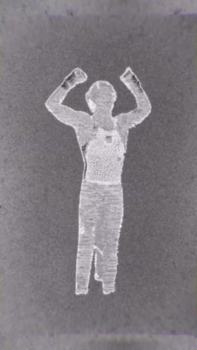
Epke Zonderland - 'Hij staat'
Top performance on the nanoscale
In these stop-motion videos, you see a fragment of the Olympics divided into about one hundred frames. These hundred frames are not normal pictures, but have been ‘printed’ on a 5 by 5 millimetre chip thanks to nanotechnology. So, the athlete in the video is a lot smaller than in real life: a whopping 100 micrometres in size. That's as small as the thickness of your hair or a sheet of paper!
How does it work?
After an elite athlete crosses the finish line, the video of the athlete at the finish line is cut out using software and converted into images made up of pixels measuring 300 by 300 nanometres. These images are then actually created on a chip in a lab.
The image of the top athlete on the plate then goes under an electron microscope. With this microscope, you don't use a light source and your eyes, but again use electrons to ‘see’. With this microscope, we can zoom in further on the images and get a good look at our athletes. A picture is taken of each picture. If you then put everything together, it creates a stop-motion video from the original video clip of the Olympic Games. But on a microscopic scale!
Keep an eye on this page and the Nanolympics social media channels for all the coming videos!