In order to accurately simulate treatment and predict treatment outcome, the biomechanical model has to be personalized to a patient-specific situation. Imaging modalities such as CT and MRI are ideal for personalization of the model as they record the patient-specific anatomy. Also, imaging is already routinely used in the clinic for diagnosis and staging.
However, current imaging methods are unable to determine the muscle architecture of the tongue. The muscle architecture of the tongue is essential for personalized biomechanical modeling as the precise muscle anatomy differs from patient to patient, especially when a tumor invades muscles of the tongue. Therefore, we have developed an MRI-based method for measuring the muscle architecture of the tongue.
Philips 3T MRI
Diffusion MRI
Magnetic resonance imaging (MRI) allows us to measure the diffusion of water in vivo and non-invasively. In muscle tissue, diffusion is restricted more perpendicular to muscle fibers than along fibers. Using this principle, we are able to reconstruct the muscular architecture of the tongue.
Commonly, diffusion tensor imaging (DTI) is used to reconstruct muscle architecture. We have improved the reconstruction by using state-of-the-art algorithms to correct for motion artifacts,
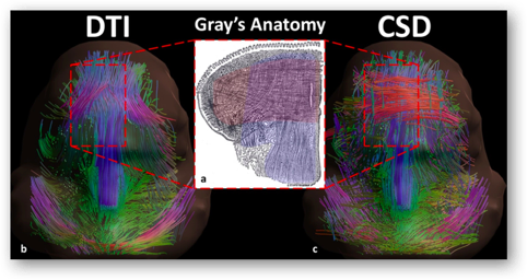
Magnetic field inhomogeneities, and crossing fibers (CSD).
Fiber reconstructions using DTI and of the state-of-the-art CSD method. Fibers are colored according to the fiber direction: red is right-left, green is anterior-posterior, blue is foot-head.
Currently, we are developing a method for automatically integrating the muscle architecture in the tongue. For more information contact Luuk Voskuilen.