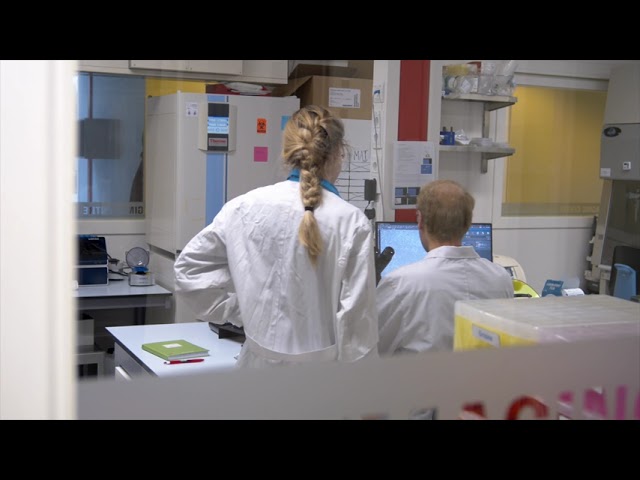
Welcome to the BioImaging Centre
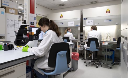
The BIC is a shared ML-I facility for advanced light microscopy at the University of Twente, featuring state-of-the art commercial microscopes, experimental microscopy techniques, image analysis software and ML-II cell- and tissue culture.
BIC Portfolio
- Commercial microscopes
- Laser scanning confocal fluorescence
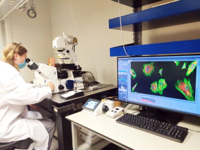
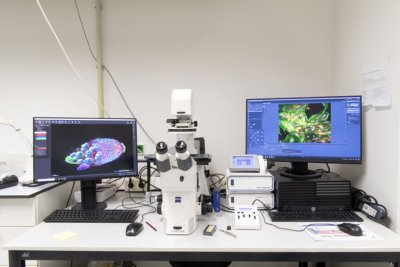
- Zeiss LSM880
The Zeiss LSM880 confocal microscope is a powerful imaging tool used in biological and materials science research. It utilizes laser scanning technology to generate high-resolution, three-dimensional images of samples, allowing for the visualization of subcellular structures and fine details of tissues and materials. The LSM880 confocal microscope can be used for a wide range of applications, including fluorescence imaging, live-cell imaging and spectral imaging.
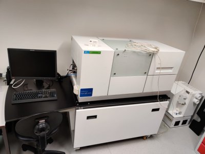
- Opera spinning disk
The Opera spinning disk is an advanced imaging system that utilizes a spinning disk confocal microscope to capture high-resolution images of biological samples.
This system is capable of high-speed, multicolor imaging of live cells and tissues, allowing researchers to study dynamic processes such as cell division and migration.
- Brightfield / Widefield fluorescence
- Zeiss Axio Observer
The Zeiss Axio Observer is a versatile microscope that can perform a range of basic imaging techniques such as brightfield, darkfield, phase contrast, and widefield fluorescence microscopy. This system is ideal for users who want to quickly visualize their samples.
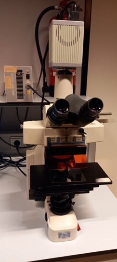
- Nicon Eclipse E400
The Nikon Eclipse E400 microscope is suitable for biological and materials science applications. It provides detailed imaging of samples and features advanced capabilities such as phase contrast and fluorescence microscopy for studying live cells and tissues.
The microscope has a camera connected to capture your images, very usefull together with our available Imaris image analysis software.
Overall, the Nikon Eclipse E400 is a versatile tool for scientific research for a variety of applications.
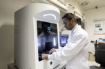
- Holotomography
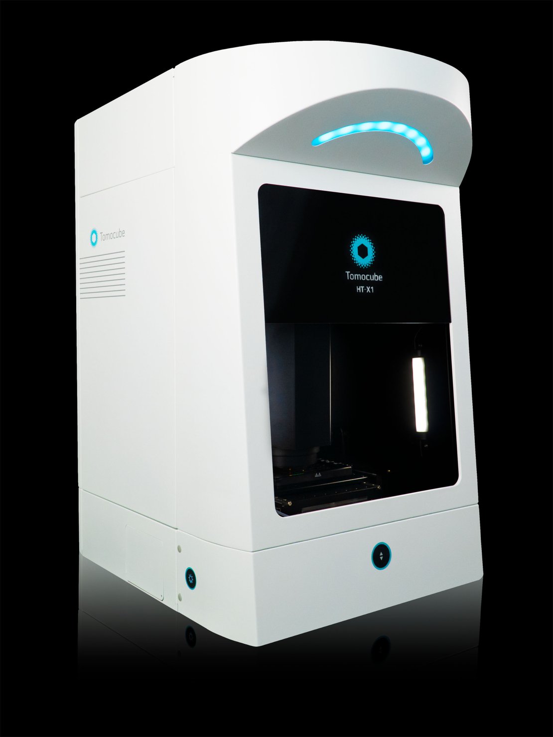
Tomocube
Observations of cellular morphology and activity often rely on labeling target molecules which can be invasive, alter the target molecules, and lack quantitative information. Common imaging tools, such as laser-based microscopes, may also cause phototoxic damage to cells. Holotomography (HT) imaging can visualize cells without labeling by capturing the light scattering properties of cellular materials using low levels of light intensity. The Tomocube HT-X1 Holotomography can collect and selectively color refractive index information to reveal cells and their organelles, while also providing quantitative 3D data such as volume, surface area, and dry mass.
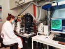
- Raman
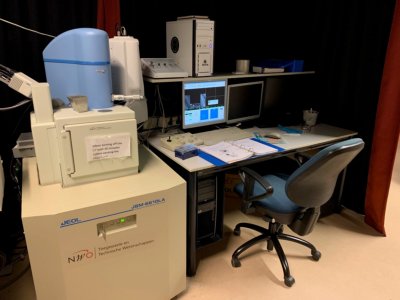
- SEM-Raman (785nm)
The SEM-Raman microscope is a powerful analytical tool that combines two distinct imaging techniques - scanning electron microscopy (SEM) and Raman spectroscopy (at 785nm)
SEM provides high-resolution imaging of sample surfaces, while Raman spectroscopy provides detailed chemical information about the sample composition.
This combination enables researchers to study the morphology and composition of materials with a high degree of accuracy, making it useful for applications such as materials science and nanotechnology research.
Furthermore, the SEM-Raman microscope can be used to study a wide range of materials, including biological samples, polymers, semiconductors, and more. Overall, it is a valuable tool for characterizing the properties of complex materials and investigating their structure-function relationships.
- Raman (640nm)
- Raman (405nm)
- Witec alpha 300 Raman (532, 633, 785nm)
- Experimental microscopy
Next to commercial microscopes we also work on novel microscopy techniques to visualize even more!
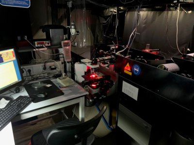
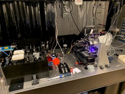
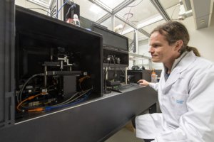
- Wavefront-shaping 2-photon microscope
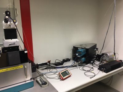
Our homebuilt Two-Photon laser scanning microscope is a powerful imaging tool used in biological and neuroscience research. It uses high-intensity infrared laser pulses to excite fluorescent molecules in living tissues, allowing for the visualization of fine details deep within the sample. Compared to traditional confocal microscopes, it can image deeper and with less photodamage. This microscope is ideal for studying the structure and function of living tissues, including the brain, and organs on a chip systems.
Additionaly our Two-Photon laser scanning microscope includes a wavefront shaping module which can correct for tissue distortions and sample abberations which allows for deeper imaging of biological samples. This module enhances the microscope's capabilities, enabling researchers to visualize previously inaccessible structures and processes within living tissues.
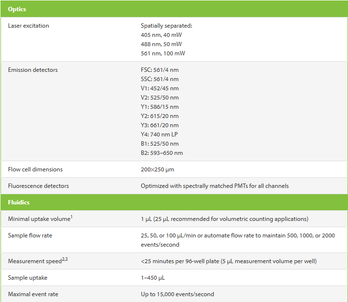
- Flowcytometry
MILTENYI VYB ANALYZER
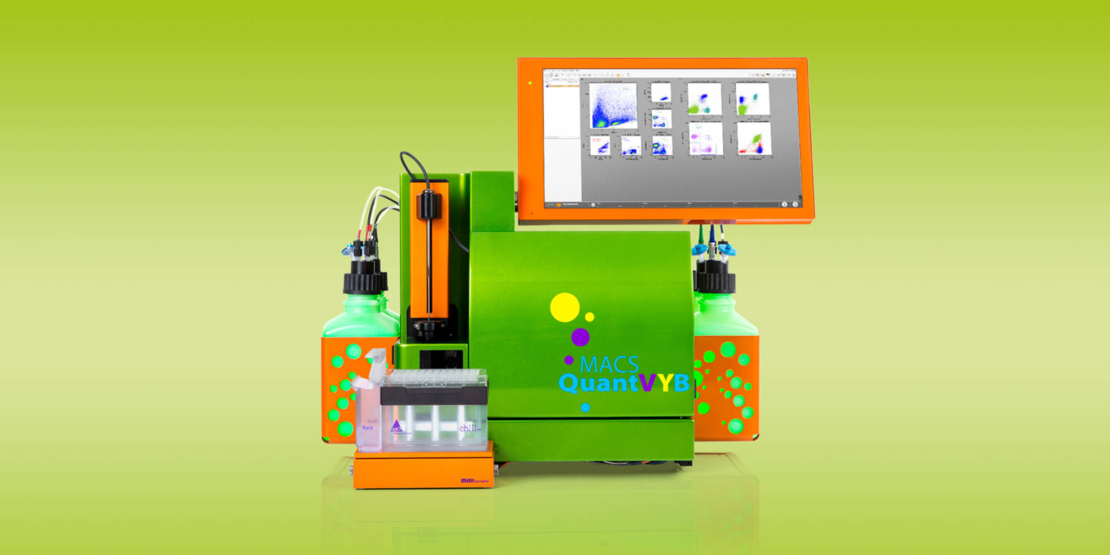
The Miltenyi VYB analyzer is all about performance, automation, compactness, and convenience. It combines all these features, but also comes with a uniquely configured optical bench, featuring violet, yellow, and blue lasers. It is a perfect match for users utilizing fluorescent protein reporters or for users that want to simply use PE and FITC conjugates.
- Biological Sample facilities
- ML - I
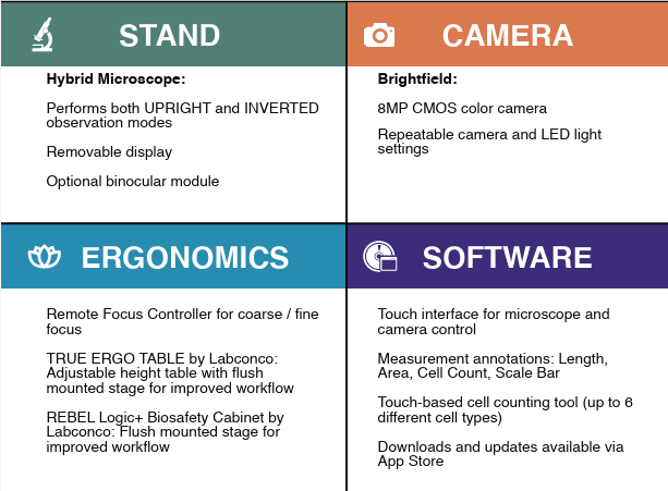
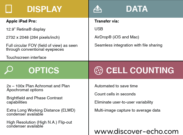
- Imaging inside Laminar Flowhood

Imaging your cells inside a laminar flowhood has never been more easy, with the Echo Rebel. Simply rotate the microscope to use it as an inverted or upright microscope for well plates, microscope slides or other cell/tissue holders.
- Image analysis software
The BIC presents IMARIS as image analysis software for data analysis free of charge to all of our users. The software can be used on the stand alone Imaris PC in the BIC lab (ZH161) or via Remote Desktop access.
If you want to make use of IMARIS, please send an E-mail to: TNW-BIC-Support@utwente.nl
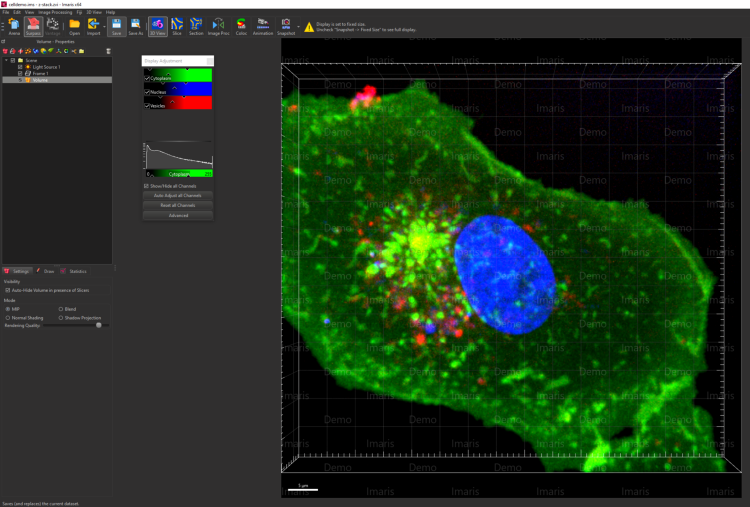
We also offer help with other image analysis software like ImageJ, FIJI, MicroManager.
Our goal is to assist researchers in addressing complex imaging challenges, providing training and support to users of our facility, to ensure that users have the skills and knowledge needed to operate the equipment effectively.
In addition, our technicians are available to troubleshoot issues and provide guidance on experimental design. For researchers requiring more advanced imaging solutions, our technicians can also work with them to develop customized imaging protocols and techniques.
The BIC is available for both University of Twente users and external users.
HOw to use the BIC facility
In order to request a device introduction and make use of the BioImagingCentre facility, we kindly ask you to fill in the following ONLINE FORM.
In this way we have all necessary information and we will send you a proposal for a date and time to introduce you to one of our systems and facility.
The BioImaging Centre is a TechMed facility and affiliated with the NL-BioImaging AM distributed infrastructure.