A SQUID (Superconducting Quantum Interference Device) is an extremely sensitive sensor for magnetic flux. Using micro-fabricated SQUID sensors, the Scanning SQUID Microscope presents a powerful scanning probe technique to locally image magnetic fields. We use scanning SQUID microscopy for various studies, such as on the occurrence of ferro-magnetism in novel thin film materials and heterostructures and for the study of fractional vortices occurring in p-phase shift structures employing the d-wave symmetry of the high-Tc superconducting state.
- Magnetic imaging using scanning SQUID microscopy
Superconducting quantum interference devices (SQUIDs) are the most sensitive magnetic flux sensors. The Scanning SQUID Microscope is a powerful scanning probe technique and uses a SQUID to locally image magnetic fields. Using conventional planar SQUID chips our microscope has been used to study ferromagnetism in magnetic oxides (see right panel Fig. 1), the study of fractional vortices in pi-phase shift structures, and the d-wave order symmetry in high-Tc superconductors.
Recent work has focused on the improvement of the SQUID probes by taking inspiration from other scanning probe techniques and integrating a focused-ion-beam patterned SQUID on an atomic force microscopy cantilever (see left panel Fig. 1). Integrating SQUID probes on conventional AFM scanning cantilevers allows for imaging at true nanoscale resolution and scanning in close proximity to the sample. Furthermore, by combing scanning probe techniques multi-modal imaging experiments by simultaneous imaging of topography, magnetometry, and thermometry.
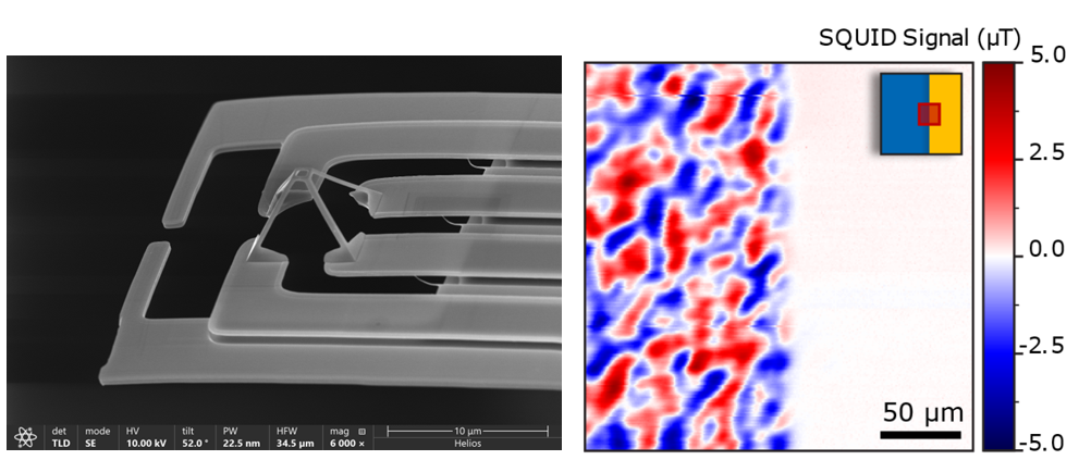
Figure 1: Left; SEM image of a nanoscale SQUID-on-cantilever probe. Right; Scanning SQUID microscopy image of partially Ti-covered LaMnO3 film showing the suppression of ferromagnetism in the covered area [1].
Are you interested scanning SQUID microscopy? Do not hesitate to contact us:
Selected recent & key publications
B. Folkers, T. Jansen, T.J. Roskamp, P. Reith, A. Timmermans, D. Jannis, N. Gauquelin, J. Verbeeck, H. Hilgenkamp, and C. M. M. Rosàrio, “Imaging the suppression of ferromagnetism in LaMnO3 by metallic overlayers”, Physical Review Materials 8 (2024), 054408.
M.I. Faley, P. Reith, C. D. Satrya, V.S. Stolyarov, B. Folkers, A.A. Golubov, H. Hilgenkamp and R.E. Dunin-Borkowski, “MoRe/YBCO Josephson Junctions and pi-loops”, Superconductor Science and Technology 33 (2020), 044005.
H. Boschker, T. Harada, T. Asaba, R. Ashoori, A.V. Boris, H. Hilgenkamp, C.R. Hughes, M.E. Holtz, L. Li, D.A. Muller, H. Nair, P. Reith, X. Renshaw Wang, D.G. Schlom, A. Soukiassian, and J. Mannhart, “Ferromagnetism and conductivity in atomically thin SrRuO3”, Physical Review X 9 (2019), 011027.
L.V. de Groot, K. Fabian, A. Béguin, P. Reith, A. Barnhoorn, and H. Hilgenkamp, “Determining individual particle magnetizations in assemblages of micro‐grains”, Geophysical Research Letters 45 (2018), 2995-3000.
P. Reith, X. Renshaw Wang, and H. Hilgenkamp, “Analysing magnetism using scanning SQUID microscopy”, Review of Scientific Instruments 88 (2017), 123706.
X. Renshaw Wang, C. J. Li, W. M. Lü, T. R. Paudel, D. P. Leusink, M. Hoek, N. Poccia, A. Vailionis, T. Venkatesan, J. M. D. Coey, E. Y. Tsymbal, Ariando, and H. Hilgenkamp,” Imaging and control of ferromagnetism in LaMnO3/SrTiO3 heterostructures”, Science 349 (2015), 716-719.